Ideas and discoveries
Researcher Jakub Lázňovský is developing software for objective evaluation of vertebral fusion success
He can read computer images. He cuts through them, examines their layers and sees the necessary parameters, which he can write into algorithms to meet the requirements of "customers", whether they be scientific colleagues or commercial clients. Jakub Lázňovský from CEITEC BUT is working on computer image processing. In one of his recent publications in the scientific journal Computers in Biology and Medicine, he presented automated software that allows objective and accurate assessment of the success of vertebral fusion of the spine of pigs in 3D. This innovative methodology has the potential to influence clinical practice and, in the future, help physicians determine the quality of fusion and evaluate the success of treatment for patients with spinal problems.
You got into software development almost by accident, is that true?
Basically, yes, because I got involved in it by collaborating with Associate Professor Vojtová's research group, which develops new materials for biological applications. They were specifically interested in intervertebral fusion, which is a surgical procedure that is used to treat a wide range of spinal conditions, such as back pain due to degenerative diseases, injuries, instability or spinal deformity. During this surgery, two or more vertebrae are fused together using implants and screws to prevent movement between them. This relieves the patient of both the symptoms of the disease and the pain.
And your colleagues have come up with a new type of implant?
That's right. During surgery, the affected disc between two vertebrae is usually removed and then graft material is inserted into the area to support bone growth. This creates a connection between the two vertebrae, which gradually fuses together to form a stable bone bridge. The bone graft is most often taken directly from the patient – from the iliac crest. Associate Professor Vojtova's research group has developed two types of new polymer/ceramic foam-based nanocomposite implants incorporating either growth factors or polyphosphates. This new type of nanocomposite implant can be used to support vertebral growth instead of graft material, the removal of which is another challenging intervention for the patient, prolonging their recovery.
What was your role?
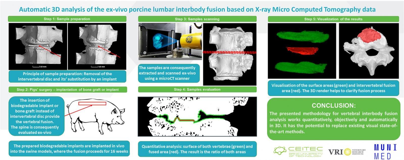
My role was to characterise the bone fusion – to see how good the fusion was and to compare the results using different types of implants and bone graft. To do this, I designed a methodology with an automated/objective approach to assess spinal fusion in 3D using X-ray computed microtomography (microCT). This is because the success of fusion is often assessed subjectively – it depends on the experience of the radiologist. However, I must emphasize that our model was not a human spine, but a pig spine, which is most similar to the human spine.
Could you describe the research procedure for a better understanding?
First, three groups of four-month-old pigs weighing approximately 40 kg had to undergo surgery on their lumbar spine. Each group received a different implant – a graft from their own bone and the two new biomaterials mentioned earlier. For 16 weeks, the aforementioned fusion, vertebral healing was in progress. Then the samples were extracted. All the spine pieces from a total of 15 pigs were scanned using microCT and I started working on the automated evaluation of the results.
What's the advantage of microCT?
It is a method that allows us to visualize the internal structure of the scanned object in 3D relatively quickly and inexpensively. Very detailed, non-invasive and non-destructive. Therefore, microCT is also a suitable tool for this research. So, I had scans of 15 spine specimens, but lacked an objective methodology to evaluate them – whether the adhesion was of good quality or not. There are only established methodologies that serve as a guide for radiologists. They evaluate the quality of the fusion by looking only at the radiographs, which they then categorize based on their quality. The problem with this approach is that it is subjective and inaccurate. That's why I wanted the computer to do it for the human and the result – how well the vertebrae fused – to be expressed directly as a percentage. Which was done.
Who will be able to work with the automated analysis of bone growth quality?
Anyone in the world who studies materials. The whole process is automatic there is no room for any subjective evaluation and therefore misinterpretation of the results. So, anyone developing bioimplants for vertebral fusion can use our methodology for evaluation. The results of fusion using other bioimplants will then be comparable, for example, to the results obtained using bioimplants developed here at CEITEC.
Which implant from the three groups analysed helped vertebral fusion the most?
The bioimplant developed by Associate Professor Vojtová's research group achieved better results than the gold standard – using a bone graft from the iliac crest. Since this is an expensive material, the colleagues wanted to test an even cheaper adaptation. Unfortunately, the suitability of this modification for use in vertebral fusion was not confirmed. However, I cannot say much about the material, as I am not an expert. I have examined the measured data.
The publication in question was published last June. Has the research moved on?
We have received a grant from the BUT as part of internal support to further develop this methodology towards more in-depth testing of the quality of individual materials that can be used for vertebral fusion. This means that another paper has already been published and with it a software package that provides a methodology for assessing the quality of fusion. It is publicly available to all interested parties.
Where might your software ideally be used?
As the approach is universal and automated, this methodology has the potential to create a standard approach for assessing fusion quality in ex vivo specimens that can then be applied to clinical data. We would be happy to be followed up. My vision is that this would make it into clinical practice to check the success of fusion. But for that we need to modify the methodology, because microCT for research use has an order of magnitude better resolution compared to clinical – as we know from hospitals. So, then you need to assess slightly different parameters of fusion quality. So, my goal is that the software will one day help radiologists in determining the quality and evaluating whether the surgery was successful.
This is your third year studying for your PhD at CEITEC, how broad is your focus?
Very broad. Our research group led by Professor Kaiser, or rather the CT lab led by Associate Professor Zikmund, works on both pure research and industrial projects. I personally study the internal structure of biological and industrial materials. In addition, we are working on how to improve the imaging of difficult-to-measure materials. For example, biological samples are harder to image because of the small difference in contrast between the different types of soft tissue in the sample. In CT, the rule of thumb is: the greater the difference in density between the different materials being imaged, the better the contrast between them. I'm sure you know this from X-rays. You can tell that the patient has screws in the bone – they glow a bright white colour. And of course, we're also developing the methodology, the algorithms, the aforementioned software to process the data.
So, you're often asked by colleagues to characterise the material? Even companies?
Yes. That was actually the case with the publication we're talking about. And we also focus on working with industrial companies. It's common that a company – a car company, for example – sends us a product and we do a quality control. We look at porosity, manufacturing accuracy, any defects...
What's the most interesting thing you've got in the lab?
I think it was a meteorite that fell near Žďár nad Sázavou. We've also studied the development of mouse embryos, nanomaterials, batteries, pharmaceuticals and even an Olympic helmet. All projects are very unique and interesting. Then I also worked with clinical data from hospitals, where the goal was to optimize respiratory support for premature babies. I basically modelled their respiratory system based on this data. So, I mostly focus on those biomedical topics with a possible overlap of improving the quality of human life.
Source: CEITEC BUT
A priceless offer. CEITEC will help researchers to bring the results of their work to the market
“I expected chaos, dirt and mess,“ says a researcher from CEITEC BUT who taught children in the Himalayas
A thin surface on the phone instead of four lenses. Metasurface will help us analyse our lunches one day
Gene therapy in developing countries. Alžběta Ressnerová is working on an easier application of CRISPR-Cas9
100 years since the birth of Josef Dadok, pioneer of instrumentation technology