Ideas and discoveries
Mother’s protein intake during pregnancy can affect newborn’s facial development
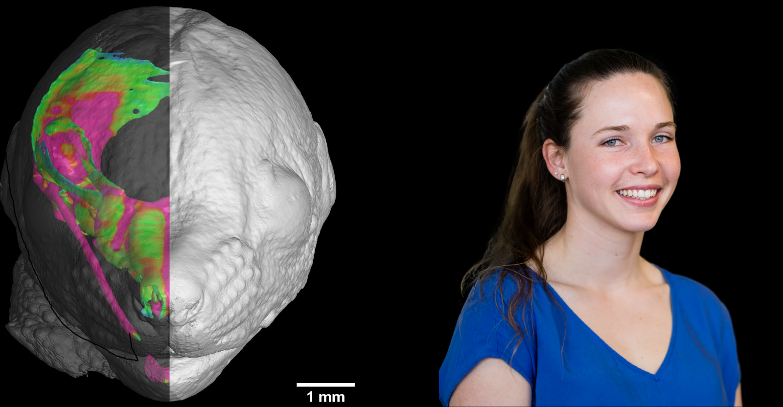
A research team from CEITEC BUT led by Markéta Kaiser participated in an international study to investigate the effect of protein intake on the fetus during pregnancy. The study was conducted on mice, or mouse embryos to be more precise. Thanks to the imaging possibilities offered by computed tomography in our CT Lab CEITEC, it was possible to create illustrative 3D models of cartilaginous tissue that clearly showed the differences between mouse embryos with different protein intakes.
At CT Lab CEITEC, researchers led by Prof. Jozef Kaiser focus on basic and applied research in collaboration with industry. The industrial partners are critical in applied research. Computed tomography is a non-invasive, non-destructive method that allows the detection of cracks or other defects on a wide variety of components so that if a company discovers that there is a problem somewhere, the CT Lab staff can detect and describe the problem without the company having to cut up or otherwise dismember the expensive product or component. The knowledge gained in this way in turn advances the researchers’ understanding of the phenomenon.
When it comes to basic research, the researchers are involved in so-called reverse engineering, where professional data display and analysis programs, which are primarily designed for mechanical engineering, are used in biology. Using advanced methods, they are able to image not only bones but also soft tissues such as cartilage or muscles.
This is also the subject of the recently published study “The level of protein in the maternal murine diet modulates the facial appearance of the offspring via mTORC1 signaling”, which appeared in prestigious Nature Communications.
Scientists from the Laboratory of X-ray Computational Micro and Nanotomography at CEITEC, led by Markéta Kaiser, have been collaborating on this study since 2018. The multidisciplinary international team was coordinated by prof. Andrei Chagin from the University of Gothenburg, Sweden. “Since then, dozens of samples have been analysed in our laboratory. Our task was not only to create 3D models of cartilage tissue, but also to analyze these 3D models. Here, we have the advantage of being a technical university and working closely with industry in the lab, and so we are using just industrial tools in biological research in a somewhat unconventional way, which is not quite so common and can bring interesting insights into these interdisciplinary studies,” says Markéta Kaiser.
We now know that facial shape and features in mammals are influenced not only by genetics but also by the mother’s lifestyle during pregnancy. It is known that, for example, the consumption of alcohol or other addictive substances can lead to facial abnormalities or developmental disorders. The present study reveals a possible new link between the offspring’s face and pregnancy lifestyle, specifically protein intake during pregnancy.
The study was conducted on mice or mouse embryos. “Specifically, the researchers focused on the mTORC1 signaling pathway, and whether protein content in pregnancy diet affects mTORC1 and plays a role in facial bone structure. Pregnant mice were fed a low- and high-protein diets, then the effect on the aforementioned signalling pathway was investigated and facial development in mouse embryos was compared,“ Markéta Kaiser explains the methodology.
It was at this point that X-ray computed tomography analysis played a key role. Scientists from CEITEC BUT created 3D models of the cartilaginous tissue that is the basis for the subsequent development of the face. The differences between embryos from mice having different protein diets were noticeable, albeit very subtle. For example, an enlarged nasal cavity or an elongated jaw were observed. Therefore, the study shows that the amount of protein in the diet can affect mTORC1 and thus the subsequent musculoskeletal development of the individual.
This collaboration certainly does not end with the published article. The researchers already have more projects planned that could help unravel other processes related to more than just facial development.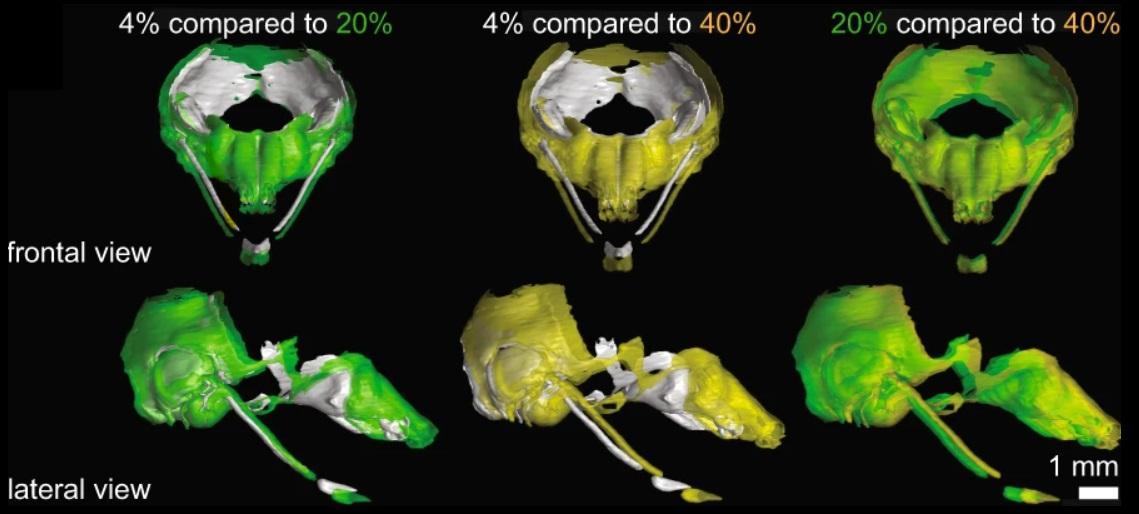
Source: CEITEC BUT
Brno is an oasis of peace for me, I feel relaxed here, says Chinese doctoral student Xia
Researchers are discovering the hidden possibilities of industry and setting a new direction in biology. In the brand new CT laboratory
In the Footsteps of the Invisible or Electron Microscopy in Brno
Awarded student Hana Kopřivová aims to come up with a new system for diagnosing tumors
We need women in science, says Forbes award-winning scientist Michaela Vojníková