Topic
In the Footsteps of the Invisible or Electron Microscopy in Brno
In the vast universe of scientific research, there is an area with such a small scale that it escapes observation by the human eye. It is these tiny particles and structures that we can explore with electron microscopy, a field that has revolutionised our understanding of the world. With its unrivalled ability to visualise structures at nanometre scales, electron microscopy has broken new ground in disciplines ranging from biology to materials science. We use the full spectrum of applications here at CEITEC, where our research simply cannot be done without electron microscopes. Since Brno is the imaginary queen of electron microscopy, we have the opportunity to participate directly in the development of this technology and to be part of the innovation ecosystem, the fruits of which can be observed during events such as the Electron Microscopy Days.
Electron Microscopy Days
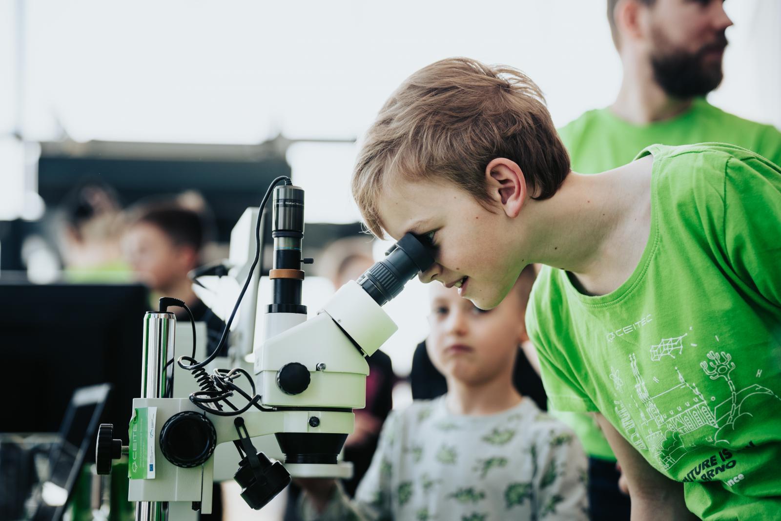
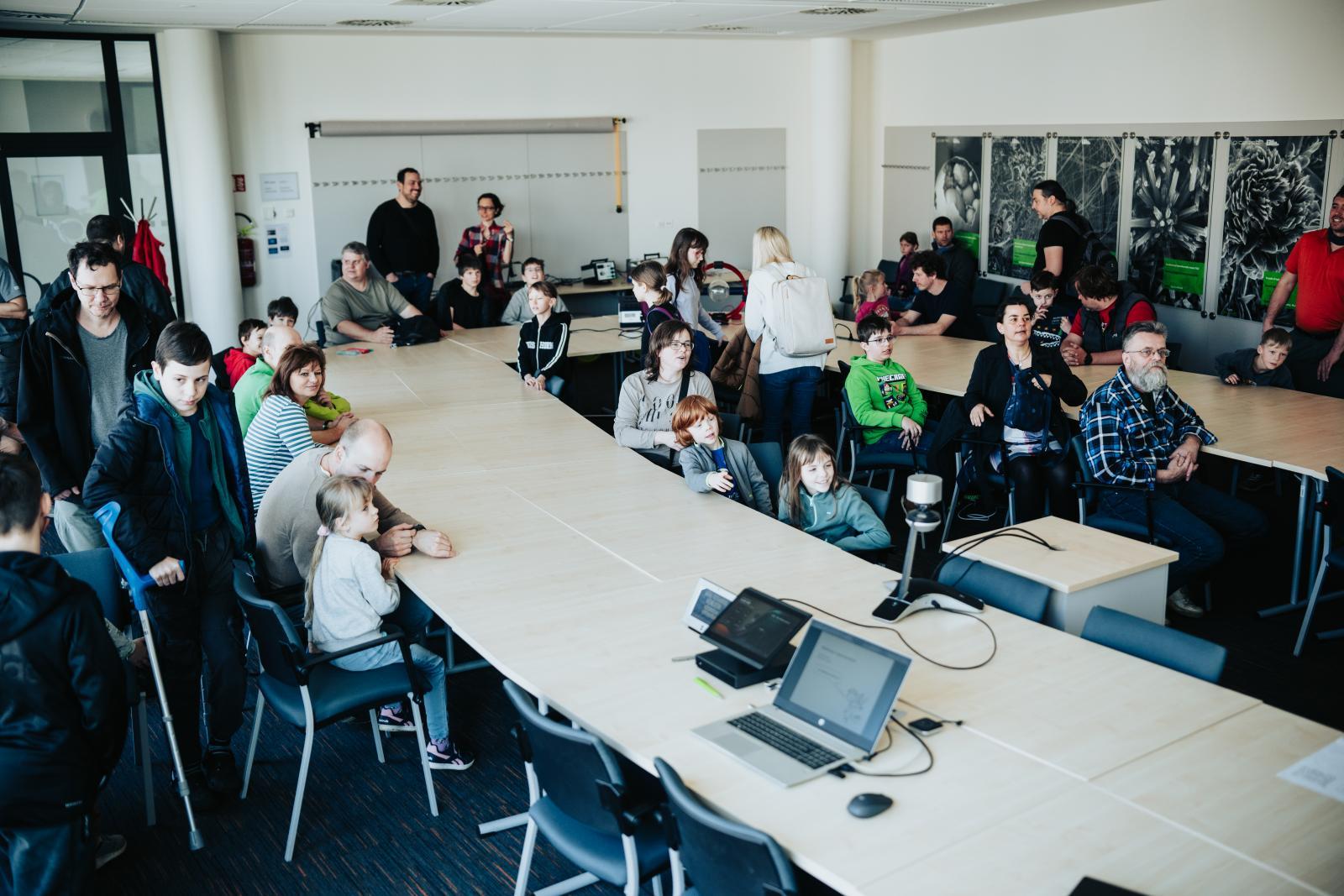
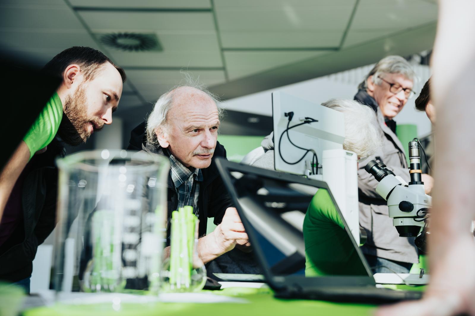
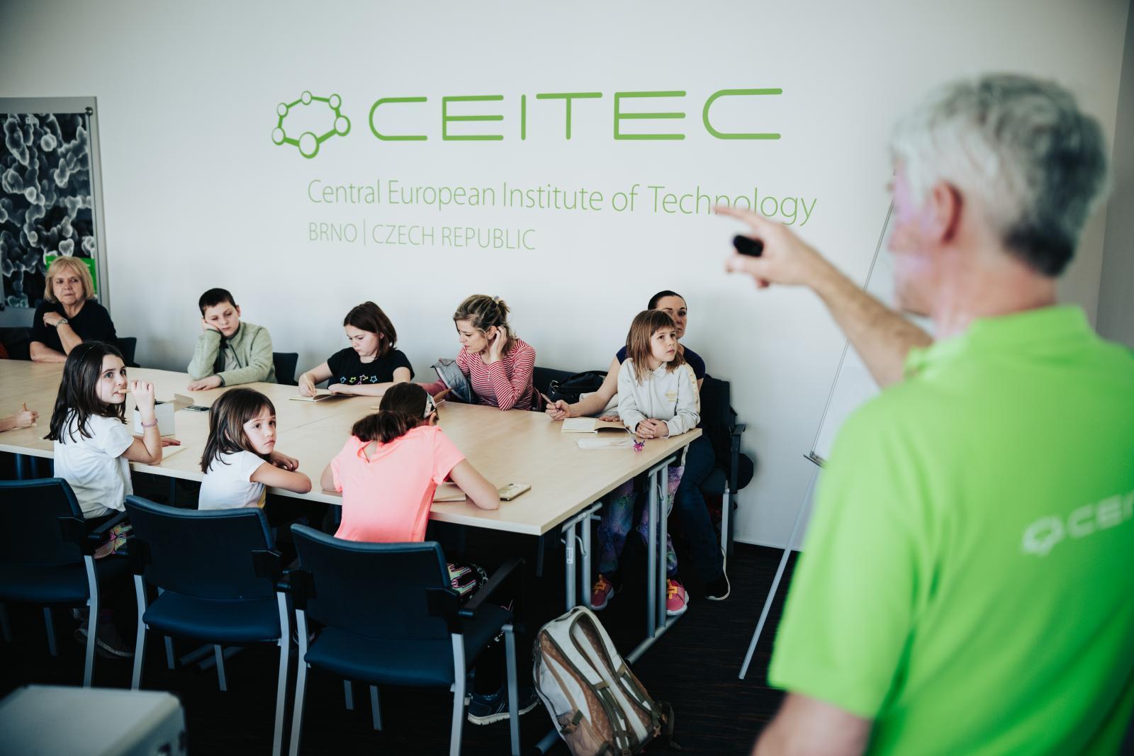
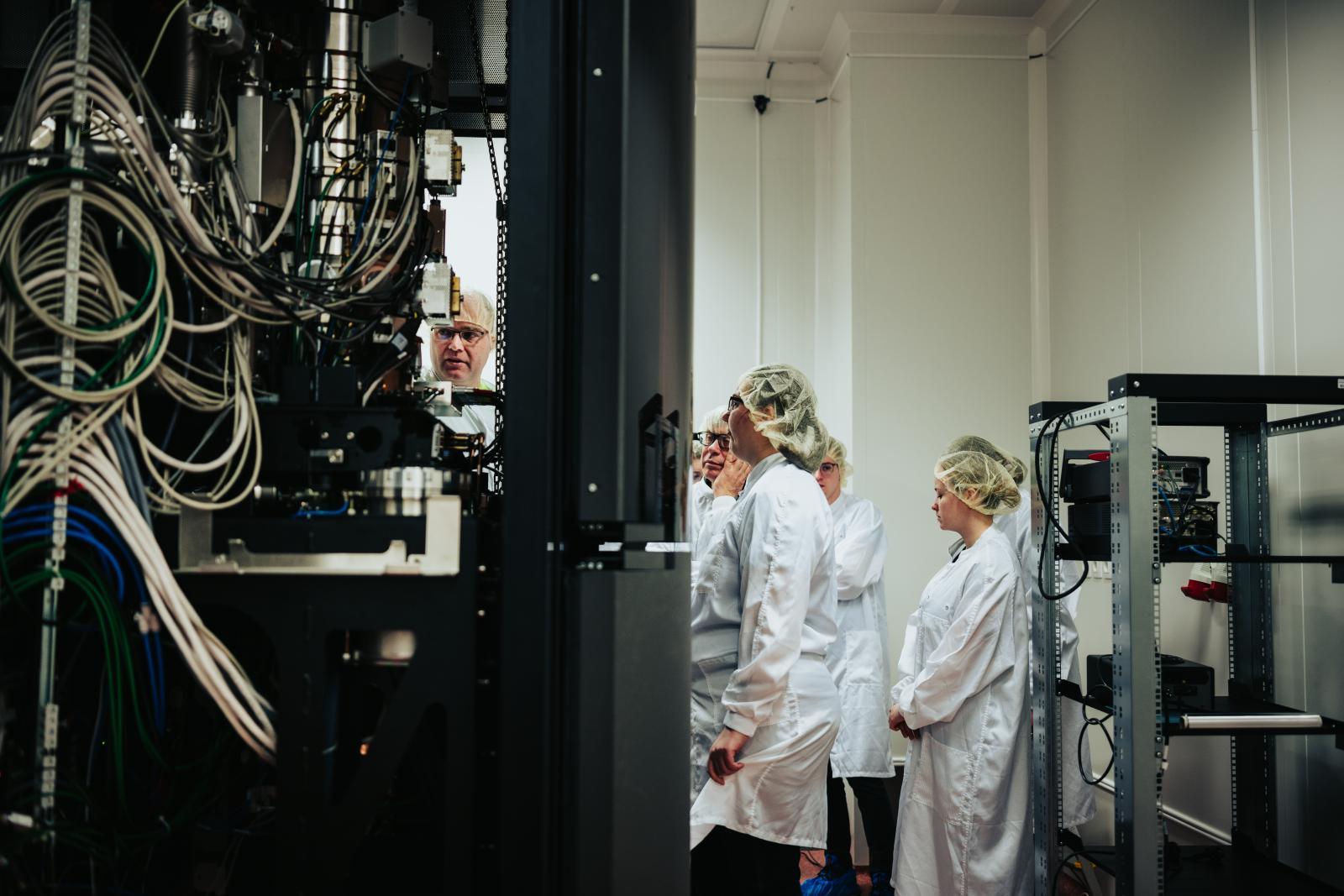
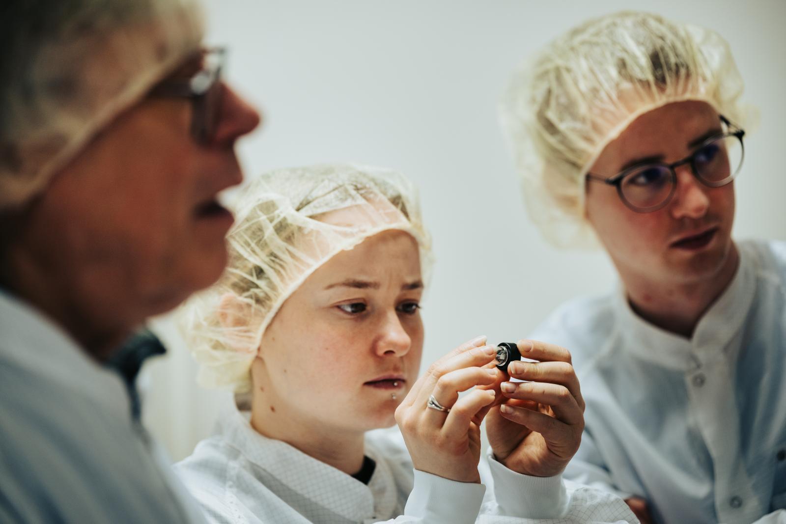
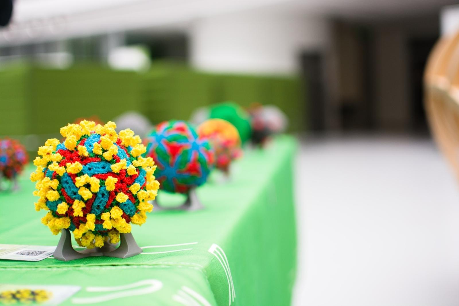
In March, CEITEC traditionally joined the Electron Microscopy Days. Both our campuses in the week of 18-23 March 2024 came alive in the spirit of this popularization event organised by Brno Observatory and Planetarium, which involves a number of partners spread throughout Brno. Every year, universities, scientific and popularization centres and manufacturing companies open their doors to the general public, especially children, and prepare rich programmes for them: from lectures to excursions to laboratories and production facilities to scientific games and experiments. The theme chosen for this year was “Villains of the Microworld”. Each institution handled the theme differently, so the main villain was the mite, the symbol of this year’s Electron Microscopy Days, but also viruses at CEITEC MUNI or electrons at CEITEC BUT. Hundreds of people visited the various lectures, exhibitions and experiments at CEITEC this year – from schoolchildren on field trips to families with children during the weekend programme.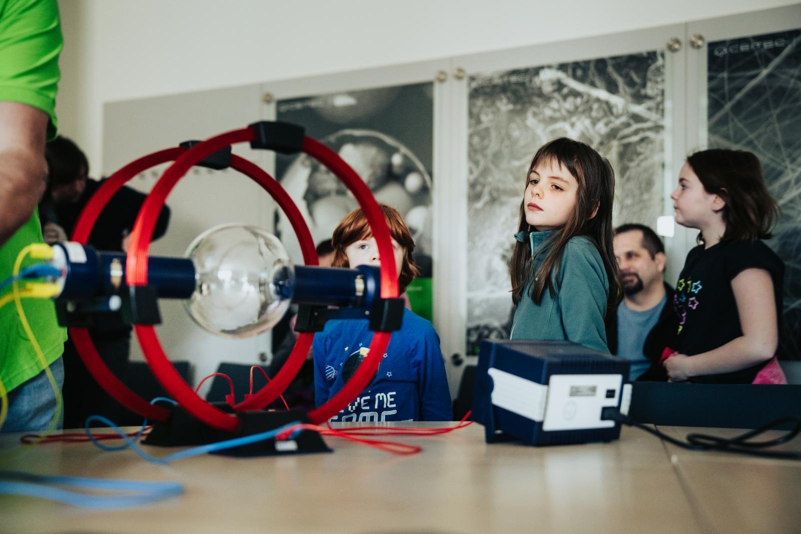
The birth of electron microscopy
Electron Microscopy Days, as the name suggests, give a glimpse under the hood of a field that is one of the cornerstones of scientific research today. The origins of this field date back to the first half of the 20th century. One of the oldest sciences, which still plays a significant role to this day – optics – was the most important for its development. At that time, it was no longer just about simple optics, the science of the propagation of light, but also about the theory of electromagnetism, in which the study of light also falls.
Determining the exact date of the birth of electron microscopy or the creator of the first electron microscope can be problematic, but what we do know is that as early as 1931 Max Knoll and Ernst Ruska in Berlin generated the first transmission electron microscope images. At that time, Soviet physicists were developing electron microscopes, too. In the following decades, electron microscopes spread all over the world and became a mainstay of scientific research in many different disciplines.
Principles of electron microscopy
Unlike conventional light microscopes, which use visible light to illuminate samples, electron microscopes use a beam of directed electrons. These electrons exhibit wave properties and have much shorter wavelengths than photons, allowing for much higher image resolution. The best electron microscopes, such as our Titans, can also image single atoms.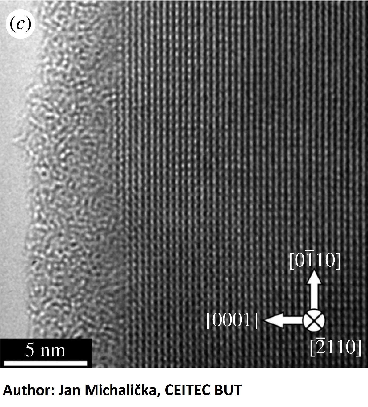
Applications across various fields
The versatile applications of electron microscopy span a variety of scientific fields and offer invaluable insights into biological, material, and environmental sciences. In biology, they enable visualization of cell organelles, protein complexes and viruses, helping us to understand fundamental biological processes and disease mechanisms. Researchers can explore tissues’ complex architecture and unlock life’s secrets at the molecular level.
In materials science, electron microscopy plays a crucial role in characterizing the relationships between the structure and properties of materials such as metals, semiconductors and polymers. By imaging the arrangement of atoms and defects, scientists can design new materials with tailored properties for applications ranging from electronics to renewable energy.
In addition, electron microscopy contributes to environmental research and protection by analysing pollutants, nanoparticles and microorganisms in environmental samples. Understanding the interactions between pollutants and ecosystems helps in designing strategies for environmental remediation and sustainability.
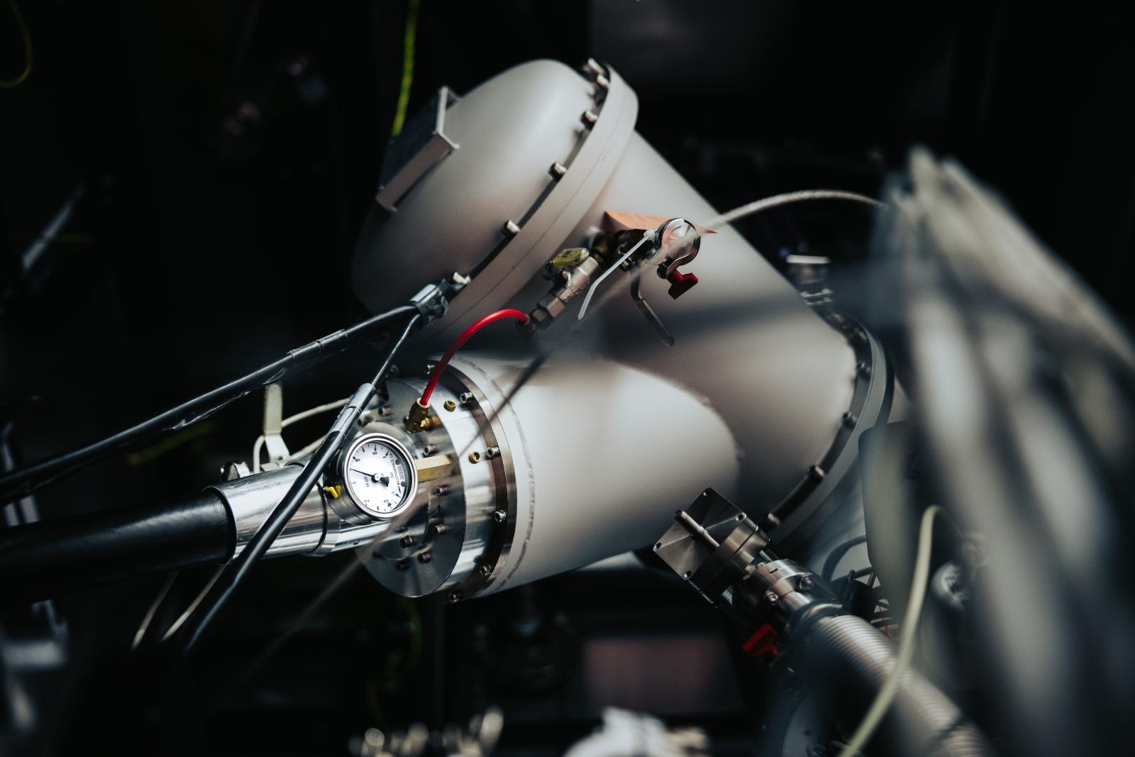
Microscopes at CEITEC
The widespread use of electron microscopes means that these instruments are essential in more or less any research and investigation we carry out at CEITEC. That is why we also have a variety of them at CEITEC, with different specifications and different resolutions – depending on what they are used for.
“The shared laboratory of cryo-electron microscopy at CEITEC MUNI uses four transmission electron microscopes, three of which have an accelerating voltage of 200 keV,” says Karel Škubník, deputy head of the laboratory. “However, the flagship of the laboratory is the Titan Krios microscope, which has an accelerating voltage of 300 keV.” At CEITEC MUNI, microscopes are used to study the structures of biological samples, such as proteins or ribosomes, i.e. cellular formations that produce proteins. Škubník also says that researchers are also looking at “viruses that infect humans or threaten entire populations. In addition, we also have three scanning electron microscopes, which are used to examine the surfaces of biological samples in particular, for instance in plants, but we have also, for example, measured the surface of a meteorite.”
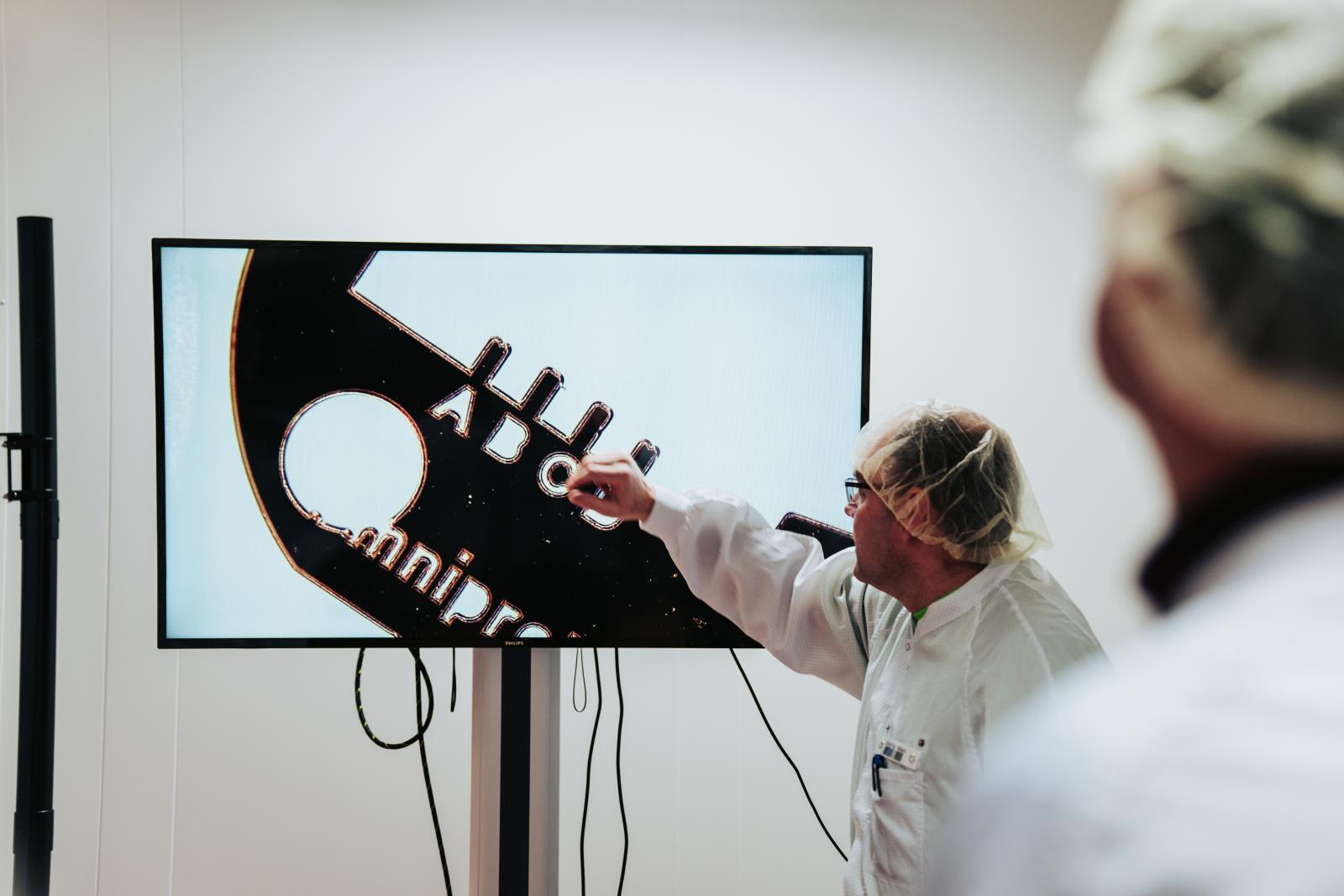
At CEITEC BUT we have seven electron microscopes, one of which is also Titan. “As these are some of the most popular instruments in our shared CEITEC Nano laboratories, which are regularly used by over 100 users, the range of applications is really wide," Lepcio explains. "The most common applications are research on advanced materials, nanoparticles, nanofibres, and other nanomaterials, as well as polymers, metals, ceramics, semiconductors, and natural materials. Microscopes serve a variety of purposes, from studying their structure, size, topography and morphology, to analyzing defects and fracture surfaces, to quality control of fabricated micro- and nanostructures.” Even at CEITEC BUT, we occasionally get our hands on more exotic samples, such as historical artefacts.
Electron microscopy in Brno
Electron microscopy is a crucial field for us at CEITEC for another reason. CEITEC is proud to be part of the Brno innovation ecosystem #brnoregion, where electron microscopy plays a vital role. With giants such as ThermoFisher Scientific, Tescan and Delong Instruments, Brno is simply the centre of electron microscopy. Every third electron microscope in the world is manufactured here!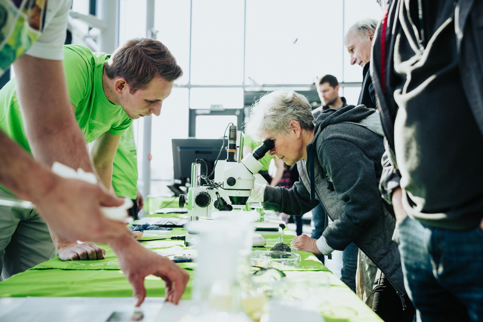
The manufacturing companies, together with research and scientific centres such as CEITEC (and other faculties at both Masaryk University and Brno University of Technology), and educational institutions such as VIDA!, form a perfectly complementary community in which researchers and former students participate in the development of microscopes, Brno companies manufacture and supply them, and here in research centres, we work with them and discover new applications. This fruitful synergy in Brno's microscopy ecosystem manifests, for example, during the Electron Microscopy Days or in the newly established Master's Programme in Microscopy, which the Faculty of Science at MUNI opens this year.
“Brno is a city of science, research and innovation. It is also a city of cooperation. It is thanks to the cooperation of companies in the field of electron microscopy, the academic sector, scientific-popularization institutions, and the public sphere that we have been hosting an absolutely unique event for several years. Its contribution is enormous – it popularizes science, promotes interest in studying technical and natural science disciplines, and represents a very promising field as well as the technological potential of our city,” concludes Brno City Mayor Markéta Vaňková.
And how did the visitors enjoy the Electron Microscopy Days programme at CEITEC? You can see for yourself in the photos below.
Author: Petra Králová
Source: CEITEC VUT
Electron microscopy is one of the key themes of the 125th anniversary of BUT.

National centre of competence for industrial 3D printing of polymer materials. CEITEC scientists to contribute their know-how
Innovative composite from TRICERA ensures lower weight of safes or higher resistance of police shields
Michaela Vojníková: Popularizing science is important. Whoever doesn't understand this is working against themselves
Researchers are discovering the hidden possibilities of industry and setting a new direction in biology. In the brand new CT laboratory
PROFiBONE: A groundbreaking material for biomedicine is being developed. It can be used to 3D-print bone replacements