At secondary school, he was fascinated by how a device as small as a mobile phone could take surprisingly high-quality photographs. Today, optics allows him to look much further – right inside living cells. Jakub Dokulil, a graduate of the BUT Faculty of Mechanical Engineering and a PhD student at the Research Institute of Molecular Pathology in Vienna, builds microscopes that can track the movement of individual molecules with nanometre resolution on millisecond timescales. He is developing a new generation of super-resolution microscopy that enables biologists to observe previously hidden processes.
When did you first become interested in optics and physics?
The first spark came back at secondary school. I wanted to undertake a secondary school research project, so I contacted Associate Professor Průša. I was curious about how we could get such high-quality photographs from a device as small as a mobile phone. He took me under his wing, let me work on the topic and gave me the opportunity to visit several laboratories. That is how I got into physics and discovered fairly early on what really interested me. I still use the fundamentals of geometrical optics I learnt back then in the laboratory today.
So when you started at BUT, you already knew which direction you wanted to take?
Exactly. That is something I really appreciate. Many people finish secondary school, start university and only then begin to work out what they actually want to do. I essentially skipped that stage of searching. When I started at BUT, I knew I wanted to pursue optics.
After completing your bachelor’s degree in physical engineering at BUT, you headed to Vienna. Why there?
During my bachelor’s degree, I already felt that I would like to study abroad. My original plan was to go to Jena in Germany, which is a major centre for optics. It has a university, Carl Zeiss and other companies and research institutions specialising in optics. But then I watched an online lecture by Stefan Hell in which he presented a method developed in his laboratory by Francisco Balzarotti. At the time, Francisco was setting up a new research group in Vienna. So I wrote to him. And that is where I ended up. Vienna also suited me because it is close to Brno. I have family and friends there, and I can visit for the weekend.
You stayed in Vienna for your PhD, too. But that was not quite so straightforward.
You might think that because I was staying in the same laboratory and working on the same project, my supervisor would automatically keep me on and I would just sign a contract. But that is not how it works.
The PhD programme is quite selective. Admissions take place twice a year, with around a thousand people applying in each round. Being an internal candidate was an advantage in my case, but I still had to go through the entire selection process.
Francisco wrote me a letter of recommendation, but as my supervisor, he was then excluded from the decision-making process concerning my application. Other people had to assess me and decide whether I was a strong enough candidate. In the end, I think thirteen of us were admitted in our selection round. External candidates also go through a matching stage, where the aim is to find the right candidate for a particular laboratory. It is not just about ability, but also about whether someone fits in with the group on a personal level and whether their research interests align.
What exactly are you working on during your PhD?
We are developing the next generation of super-resolution microscopes using the MINFLUX method. What makes these microscopes unique is, above all, their combination of spatial and temporal resolution. They can track molecular machines with nanometre spatial resolution and millisecond temporal resolution. That matters because many things in biology happen very quickly and on a very small scale. If you want to understand what is really happening inside a cell, a detailed image alone is sometimes not enough. You need to see how individual molecules move.
For example, Professor Musser’s research team in Texas used this method to describe the mechanism by which molecules are imported into the cell nucleus. They were able to track where importin binds to the nuclear pore, how long it stays there, and so on. These are precisely the kinds of observations that make these microscopes unique. In my project, I am working on a new generation of this technology. We want to build on observations like these and push their possibilities even further.
How does MINFLUX differ from electron microscopes?
Electron microscopes use electrons instead of light and have brought about an enormous revolution in the life sciences over the past 15 years. For example, they can determine the structures of proteins and other molecules at atomic resolution. What they can do today is amazing. But there is one major drawback if you want to observe dynamic processes. You have to prepare the sample, often freeze it, and place it in a vacuum. The result can be a fantastically detailed image of a structure, but the sample is dead. You cannot watch how molecules actually move within it.
And that is precisely where light microscopy still excels. I can take a living cell, put it under the microscope under ambient conditions and observe what is happening inside it. Because it is neither frozen nor in a vacuum, the molecules can move, and we can follow dynamic processes. For example, you could never make a “video” of molecules travelling from the cytoplasm into the nucleus during import using conventional electron microscopy. With this method, you can.
And how does MINFLUX compare with other light microscopy methods?
There are methods based on single-molecule localisation, for example, that can produce very detailed static images. But their temporal resolution does not match that of MINFLUX.
That is really the main difference that fascinates me about this method. It is not just about seeing something very small. We want to see how that very small something moves.
When you talk about a microscope, people might picture a ready-made instrument that you order from a supplier. But you essentially build yours from scratch. What does that process look like?
That is one of the things I absolutely love about my work. First, you need an idea of how the method should work – what you will do with the light and how it will interact with the sample.
Then you have to design the optical system, build it and develop a way to control it. Only then do you bring in the biological sample and find out whether you can obtain something new.
In the laboratory, we have a modular set of optical components from which we assemble the system. You buy mirrors, lenses, various mounts and other components, and then gradually put it all together. It is a bit like a Merkur construction set for adults. You are constantly screwing things together and adding components, and then you have to align the system. For example, you look at the light beam on a camera and try to optimise it by tilting the mirrors. At the same time, you have to write the software that controls the whole system, because some components are computer-controlled. Colleagues in Stockholm developed a Python programme called ImSwitch for controlling microscopes. I adopted it and am adapting it to my own needs.
So as physical components are added to the table, the software is being developed and new functions are being added alongside them.
And how do you know the whole system is working as it should?
First, we carry out calibration measurements. We need to verify that the method works as expected. For this, we use tools such as nanorulers based on DNA origami technology, which allows us to create precisely defined structures at the nanometre scale. Only once we are sure that the optical system works do we move on to the biological sample. Then we try to collect actual biological data and check whether we can observe what we originally set out to see.
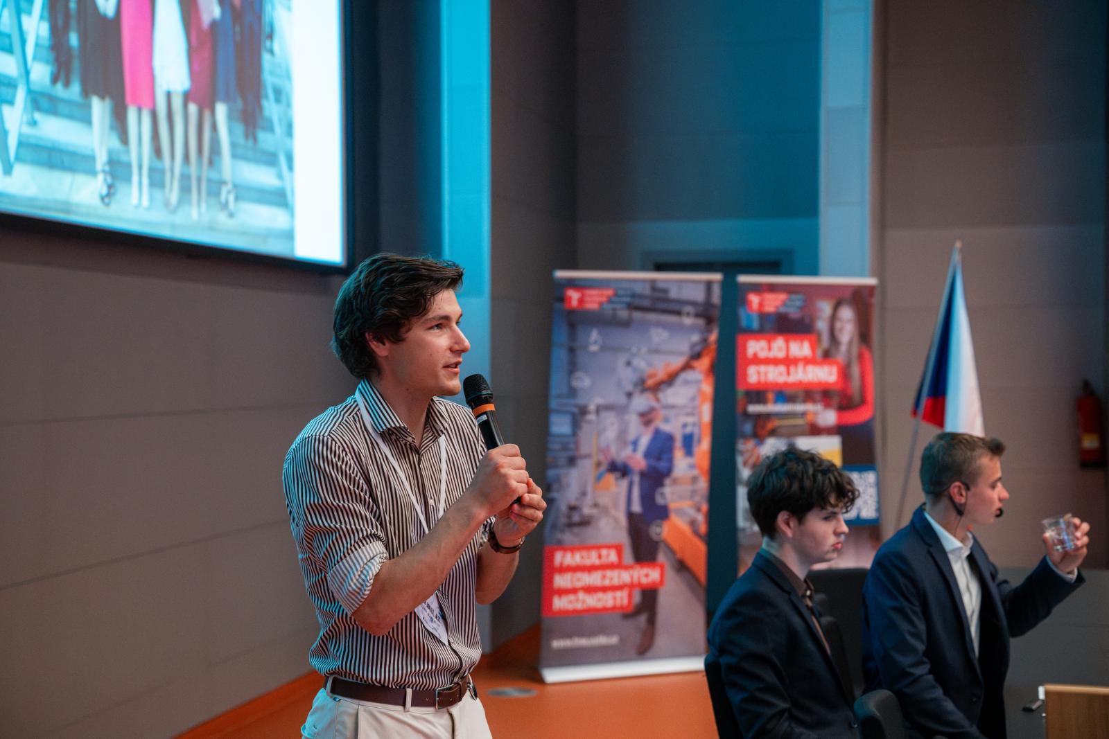
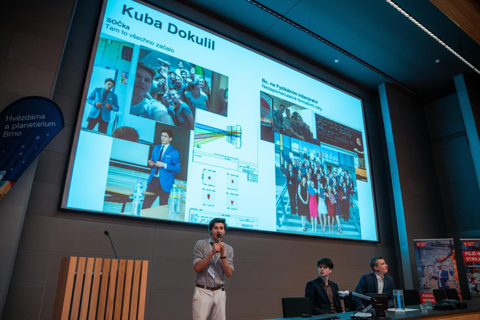
Jakub Dokulil presented his work and career in microscopy during this year's STEAM Slam at the FME BUT. The event for talented high school students is organized annually by the JCMM association. | Author: Václav Široký
What comes next?
My PhD project has two main stages. First, we want to build a new microscope that extends the capabilities of current systems. That means physically assembling it from individual components, developing the necessary software and verifying that the method works. Then we want to improve it further, particularly in terms of temporal resolution. And it is very important to me that the result does not remain a single setup on a single table in Vienna. We want as many other laboratories as possible to be able to use it.
That is why we plan to publish not only the method itself, but also the software that controls the optical setup and a practical protocol explaining how to build, configure and maintain such a microscope. If other laboratories can build and use it themselves, they will be able to obtain entirely new data. Commercialisation would, of course, be nice, but that is still a long way off and not something we are currently working on.
Ideally, where would this technology be used? In medicine, perhaps?
I understand why you ask about medicine. Our goal, though, is to develop a tool that allows biologists to see something they could not see before. So I see its application in basic research in the life sciences. We want to give biologists a tool that lets them observe new things and describe previously unknown processes in living organisms. It is certainly very possible, and indeed likely, that someone will then use those findings in medical research or to develop an application. But our primary aim is to provide biologists with a new tool. In my view, basic research is the backbone of all science.
Have you stayed in touch with BUT during your studies in Vienna?
Yes. Since I studied at the Institute of Physical Engineering, I still keep in touch with people there, particularly those in Professor Chmelík’s group.
At the moment, it is not a research collaboration as such. We worked together for a while, get on well personally and enjoy meeting up. Those connections are important to me.
You can now compare the research environments in Vienna and Brno. What are the differences?
I think CEITEC BUT offers exceptional laboratory equipment and facilities. During my master’s studies at the University of Vienna, I saw local research teams travel to Brno to use some of those instruments and facilities. I think the equipment and the range of capabilities available in one place are excellent. Vienna, meanwhile, has a very international environment. My supervisor is Argentinian, and our team of eleven includes two Austrians and a large proportion of people from abroad. The individual institutes hold open recruitment processes for research group leaders and try to attract the best people. There is a strong emphasis on quality, and researchers are very successful in securing competitive grants, including prestigious ERC grants. This creates a highly international and productive research environment.
What did BUT give you for your subsequent research career?
Above all, incredible support from the people at the Institute of Physical Engineering. Whether it was Associate Professor Kalousek, the professors who taught us or Professor Chmelík – when I said I wanted to study abroad, they supported me in every way they could. I appreciate that enormously. And I know that this kind of support is certainly not something you can take for granted in Czechia.
And where would you like to go after your PhD?
My main focus is on my PhD project and publishing the results. I am keeping an open mind about what comes afterwards. But I definitely want to put the skills I have gained during my PhD to use. That could be in academia, but equally in a high-tech company. Right now, the most important thing for me is to finish what we are working on and take our method as far as possible.
Jakub Dokulil presented his work and career in microscopy during this year's STEAM Slam at the FME BUT. | Author: Václav Široký (mar)